Table of Contents
Introduction:
Giemsa staining is a commonly used method in microscopy, particularly in the investigation of blood cells and pathogens. In addition to assisting with disease detection, it offers priceless insights into cellular architecture. We shall examine the purposes, tenets, method, and uses of Giemsa staining in this manual, as well as its benefits, and drawbacks.A Complete Guide to Giemsa Staining Understanding
Objective:
The primary objective of Giemsa staining is to enhance the visibility of cellular components, such as nuclei, cytoplasm, and parasites, under a microscope. This technique help in identifying and differentiating various cell types and potential abnormalities.
Principle | What is the Principle of Giemsa staining:
Giemsa staining is based on a combination of acidic and basic dyes. These dyes bind to specific cellular structures, imparting distinct colors. The stain works by altering the pH of cellular components, causing them to take up the dye and become visible under a microscope.
Composition Giemsa stain:
Giemsa stain play a crucial role in enhancing the visibility of cellular structures and microorganisms under a microscope.
The composition of Giemsa stain includes:
- Azure Eosin Methylene Blue: This mixture contains azure dye, eosin dye, and methylene blue dye. Azure dye binds to acidic components, staining them blue, while eosin dye stains basic components pink. Methylene blue, a basic dye, adds contrast and enhances the visibility of cell nuclei.
- Buffered Solution (pH 6.8): A buffered solution is used to adjust the pH of the stain, creating an optimal environment for dye uptake by cellular components.
- Distilled Water: Distilled water is used for dilution and rinsing steps during the staining process.
- But there are different modification of giemsa.
Depending upon the goal of study one can use different combination. Or simply used premade solution by diluting it 1/10 with water.
Procedure of Giemsa staining:
The Giemsa staining procedure involves two primary techniques: thin film staining and thick film staining.
Staining Procedure 1: Thin Film Staining:
- Prepare a thin blood smear on a glass slide and allow it to air dry.
- Fix the smear with methanol for a few minutes.
- Immerse the slide in buffered Giemsa stain for approximately 20-30 minutes.
- Rinse the slide with distilled water and allow it to air dry.
- Examine the stained smear under a microscope.
Staining Procedure 2: Thick Film Staining:
- Prepare a thick blood smear by placing a drop of blood on the slide and spreading it.
- Allow the smear to air dry and fix it with methanol.
- Apply buffered Giemsa stain to the slide for about 30-45 minutes.
- Rinse with distilled water and let the slide dry.
- View the stained slide under a microscope.
Results and Interpretation of Giemsa staining:
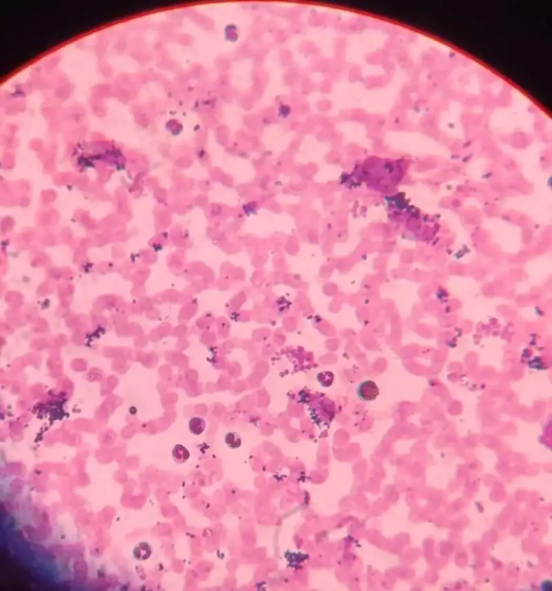
Giemsa-stained specimens reveal different cell components with distinct colors. Nuclei may appear blue-purple, while cytoplasm takes on shades of pink or pale blue. Microorganisms and parasites can also be identified based on their staining patterns.

Application of Giemsa staining:
Giemsa staining is crucial in various fields, including:
- Hematology: Identification of blood cell types and detection of abnormalities.
- Microbiology: Detection and characterization of microorganisms, such as bacteria, protozoa, and parasites.
- Pathology: Diagnosis of diseases like malaria and other blood-related disorders.
Advantages:
- Provides clear differentiation of cellular components.
- Offers rapid results and is relatively easy to perform.
- Compatible with different types of microscopes.
Limitations:
- May require expertise to accurately interpret results.
- Staining intensity can vary, affecting consistency.
- Not ideal for detecting certain subtle cellular changes.
Giemsa staining (Romanowsky-type, polychromatic stain) is a very useful in biological study, to identification of cells and microorganisms using microscope. By using a mix of dyes(Azure, Methylene blue, eosine) , it highlights different parts of cells, making them visible under a microscope. This technique is used in diagnosing diseases like malaria and studying blood. While it’s easy and quick, its accuracy depends on expertise. Giemsa staining is a useful tool in a number of scientific and health areas because it allows us to see hidden aspects in the microscopic world by revealing cellular details and microbes.
References:
Dolan, M. (2011). The role of the Giemsa stain in cytogenetics. Biotechnic & Histochemistry, 86(2), 94-97.
FAQ
what is giemsa staining used for
Giemsa staining (Romanowsky-type, polychromatic stain) is a very useful in biological study, to identification of cells and microorganisms
giemsa staining reveals which types of bands on chromosomes?
giemsa taining stains euchromatin in chromosome
what is giemsa staining ?
it is the differential straining technique in the cytological study.
what is the purpose of a staining technique of chromosomes such as giemsa
giemsa staining of chromosome gives alternated bands of light and dark on chromosome which is used to study the structural and numerical changes.
Check our other notes